Pet diagnostic testing in Dubai
Diagnostic Procedures
Pet X‑ray in Dubai
X-ray Imaging (Radiography)
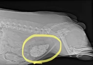
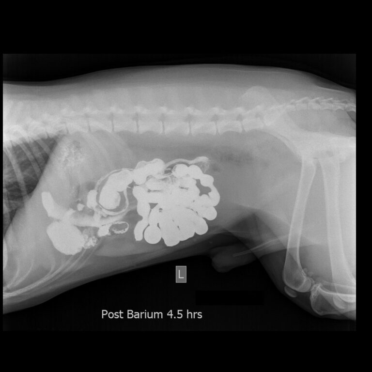
Contrast X-ray
The body’s soft tissues do not absorb x rays well and can be difficult to see using this technology alone. Specialized x ray techniques, called contrast procedures, are used to help provide more detailed images of body organs.
In these procedures the animal is given a dye that will block x rays. This can be given intravenously to examine organs like the kidneys or heart, or by mouth to examine the digestive tract. A series of x-rays is taken after the dye is given, which will outline the organs where the dye collects. This makes it easier to spot any abnormalities.
The x-ray machine is positioned so that x-rays are focused on the area to be examined. Exposure to x-rays lasts only a fraction of a second. However, the greater the exposure, the greater the risk that radiation may damage cells. For this reason, a very low dose is used, and lead shields may be used to protect areas that are not being x-rayed and the people taking the radiograph.
These images are stored digitally on computers to be shared with specialists, other clinics or the pet owners themselves.
Veterinary diagnostic lab Dubai
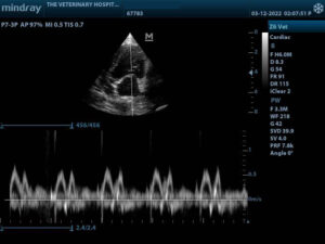
Ultrasonography (Ultrasound)
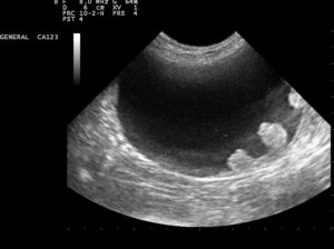
Ultrasonography (commonly called ultrasound) is the second most commonly used imaging procedure in veterinary practices. It uses ultrasonic sound waves to create images of body structures based on the pattern of echoes reflected from the tissues and organs. Ultrasound is much better than x-rays at showing the soft tissues within the body.
The veterinarian usually performs an ultrasound scan by pressing a small probe against the animal’s body, most frequently the abdominal wall. The process may involve shaving area of your pet’s hair if found in the area needed to be scanned.
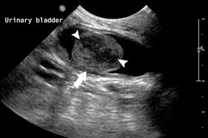
An ultrasound scan can show the size and shape of many organs and can also show abnormalities within them. An ultrasound scan is both painless and non-invasive and poses no risk of complications. Although ultrasound can be used to evaluate most soft tissues; the heart, abdominal organs and urinary bladder are the most frequently scanned in veterinary clinics.
Heart scan or Echo-cardiography is used to identify heart disease, the structure and function of the heart and its valves and for on-going optimal monitoring of the patient.
Abdominal scan is used to identify diseases and dysfunction of the alimentary canal, urogenital system and for pregnancy diagnosis.
Bladder scan is to identify urinary bladder stones (dense white mass in centre of screen).
Ultrasound can be used for examining external masses such as cancers and inflammatory problems.
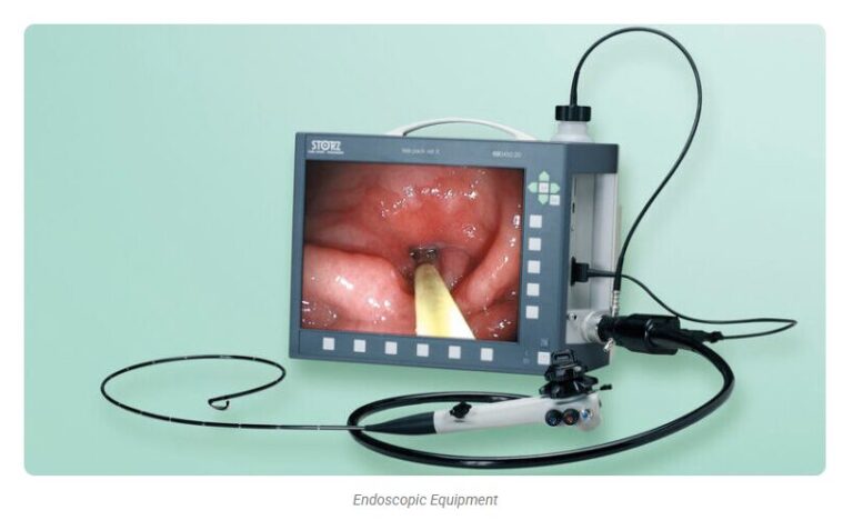
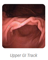
Endoscopy
Endoscopy procedures are considered non-invasive procedures and sometimes can be an alternative to surgical interventions.
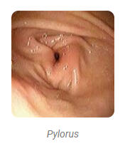
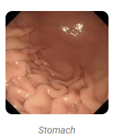
Endoscopy help veterinarians make a diagnosis of the disease that has been causing your pet’s clinical signs of vomiting, diarrhoea, weight loss, abdominal pain or swelling or loss of appetite by allowing full colour viewing of the oesophagus, stomach and the upper part of the small intestine or the colon.
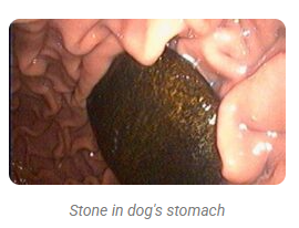
The veterinarian can identify abnormalities such as inflammation, abnormal swelling, or areas of scarring or stricture (abnormal narrowing). If a foreign body such as a bone, stick, rock, toy, coin, or hairball is seen, it can usually be seen and retrieved.
Seeing an abnormal lesion or suspicious area gives us valuable information but it is usually necessary to biopsy the area in order to reach a diagnosis. This involves taking precise biopsy samples “tiny bites” or pieces of tissue cut from the lining of the abnormal area by the biopsy instrument. These tissue samples, called “pinch biopsies” are then submitted to a veterinary pathologist for microscopic evaluation.
Many diseases cause changes that can only be detected by histopathology, or a microscopic inspection of the tissues. Therefore, even if the organ or tissues appear normal, biopsies are taken. In many cases, biopsy of the stomach of a vomiting pet or of the colon of a pet with diarrhoea will be very helpful in determining if disease is present.
The diagnosis of cancer of the gastrointestinal tract can be done by using the endoscope. However, some tumors do not affect the mucosa or inner lining of the stomach or colon. Since the biopsy procedure only samples the mucosa it is possible to miss detecting a tumour that involves only the deeper layers of the intestinal tract. In these unusual cases, the biopsy results are normal, yet the pet continues to experience clinical signs. In order to reach a diagnosis in these cases, full-thickness biopsies obtained through an exploratory surgery, (exploratory laparotomy) or non-invasive tests such as an MRI (Magnetic Resonance Imaging) may be required.
General anaesthesia is required for this procedure as it is impossible to safely pass an endoscope into a conscious dog or cat’s stomach or colon. Most pets will require only a short-acting anaesthesia and the patient is allowed to go home shortly after completion of the procedure.
Result times will vary from 3 days to two weeks, depending on the individual circumstances and which overseas reference laboratory is used.
Note:
Due to the complexity and expense of CT scan and MRI, patients requiring such diagnostic procedures will be referred to a third party facility and our veterinarians will act on the results and findings.
Reconstructive Surgery
This is generally required after severe road traffic accidents, burns and falls from high balconies. We use grafts to repair areas of significant skin loss, repair damaged eye corneas with conjunctival grafts, muscle and bone grafts in orthopaedic surgery and in thoracic and abdominal surgery.
Digestive Tract Surgery
This includes removal of foreign body blockages, stomach dilatation and volvulus (twisted stomach), gastropexy, repair of anatomical abnormalities, gallbladder surgery, splenectomy, liver lobectomy, megacolon surgery, tumour removal surgery.
Hernia Repair Surgery
Most common hernia repairs are umbilical, inguinal, diaphragmatic and perineal hernia.
Tumour and Mass Removals
From various parts of the body (internal and external masses)
Severely Damaged Organs
Eye Enucleation- removal of the eye. Total Ear Canal Ablation with Lateral Bulla Osteotomy (TECA-LBO)- removal of the complete ear canal. Nephrectomy- kidney removal. Lobectomy- liver lobe removal. Splenectomy – spleen removal.
Urogenital Surgery
Kidney/ Bladder /Urethral Surgery. Removal of kidney stones and bladder stones. Ectopic ureters.
Our Location
Villa 4, Al Wasl Rd
Umm Al Sheif – Dubai
Contact Us
Got Questions?
Emergency
ONLY hotline